The MechaMorph – may the Force be in your Images
Over decades, myology has aimed to obtain direct measures for muslce dysfunction, i.e. weakness, by using manual or tedious single fibre biomechatronics systems, e.g. to characterize maximum specific force or Ca2+ sensitivity of the conrtractile biopolymer bionsensors. Apart from immediate signaling effects on contractility, also structural changes to the sarcomeric polymer architecture during remodeling in chronic disease also sets structural limits to maximum force or biosensor response. By optimizing our multiphoton Second Harmonic Generation morphometry arrpoach to single fibre cytoarchitecture, we have repeatedly documented vast ultrastrucutral irregularities in diseased muscle fibres that may present a structural correlate to weakness. Therefore, SHG imaging can suggest weakness from disordered myofibrillar arrays.
But how to pove such a cause-effect scenario? It would require measuring force and subcellular structure at the same time in the same preparation.
In order to achieve this goal and provide a kind of ‚calibration‘ to our SHG image analysis to predict force from looking at structure, we engineered a miniaturized force transducer system that was inspired by our MyoRobot, i.e. involving voice coil technology, but would still fit between the two objectives of a multiphoton microscope for forward scattered myosin SHG imaging. Our MechaMorph (see figure) consists of two parallel troughs connected to a force transducer and a voice coil actutator pin to take up a single muscle fibre and tightly place it with polymer fibre clips.
As shown in the middle part of the figure, a single normally structured wt fibre and a disordered fibre from a dystrophic mdx mouse were analyzed regarding strucutral parameters of latice order (cosine angle sums, CAS; vernier density, VD) and recording graded pCa force responses. Lattice idsorder (low CAS, high VD) is reflected by decreased biosensor Ca2+ sensitivity (Schneidereit et al. 2018). Taking advantage of less ordered cytoarchitecture in mdx fibres the range of CAS and VD values could be expanded to areas not seen in healthy, nromally structured wt fibres. The right part of the figure shows our calibration data, confirming that fibres with less ordered structure (mdx, red; and some wt fibres) were always associated with lower maximum force force capacity.
The MechaMorph is a valuable tool to obtain calibration optics-to-biomechatronics data sets that we currently expand also to other disease models than mdx. It can even be applied to other tissues, e.g. heart strips or skin strips which will enable to obtain a ‚image-based prediction-classifier for biomechanical tissue performance‘.
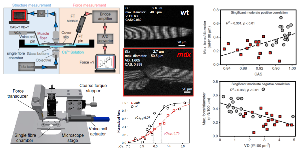
Schematics and design of the MechaMorph systems technology (left) to allow for simultaneous SHG and isometric force recordings in single fibres from healthy (wt) and dystrophic (mdx) muscle, where the former is characterized by regular ultrastructure (high CAS, low VD) and the latter by deranged sarcomere latticew in 3D (low CAS, high VD). In addition, low regularity (dystrophic) is accompanied by compromised contractile biosensor Ca2+ sensitivity. Right, direct calibration of structure-function relationship with sarcomeric regularity determining foce output (Schneidereit et al. 2018).
Reference:
Schneidereit D, Nübler S, Prlöß G, Reischl B, et al. (2018) Optical prediction of single muscle fiber force production using a combined biomechatronics and second harmonic generation imaging approach. Light Sci Appl. 7:79. doi: 10.1038/s41377-018-0080-3.