Multiphoton Endomicroscopy for Life Science and Medicine

We develop advanced endoscopy systems to implement new label-free imaging and spectroscopy techniques for in vivo imaging in a minimal invasive setting. The aims are a refinement of in vivo experiments in fundamental research and a translation of technological advancements to improved clinical diagnostics and therapy monitoring. In an FAU Emerging Fields Initiative ‚ADVENDO LIFE‚ (2014-2017), we designed and engineered a miniaturized galvanometric scanning system employing two-photon excitation through a GRIN lens optics to collect non-linear label-free Second Harmonic Generation (SHG) and Autofluorescence (AF) information of tissue architecture in pre-clinical in vivo models using small animal multiphoton endomicroscopy. The figure below shows the setup optical pathway (A), the engineered distal side-view GRIN lens optics (B) as well as in vivo endomicroscopy colonoscopic images during the time course of experimental inflammatory bowel disease (IBD) using dextrane sodium sulfate (DSS) (C). The system allows to follow microscopic changes of colon epithelium and mucosa in depth, employing an electro-tunable lens (ETL) for axial focusing in a truly minimally invasive rigid endomicroscopy setting with pulsed laser excitation.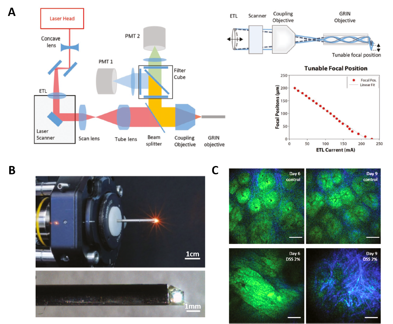 Two-photon endomicroscope ADVENDO LIFE. A, schematics of optical beampath involving galvanometric scanner, coupling objective into GRIN objective and electrotunable lens (ETL) for z-focus actuation through control of ETL current. B, images of distal optics part of final setup. C, in vivo two-photon endomicroscopy of NADH signals (green) originating from eptihelial crypt cells and Second Harmonic Generation (SHG) signal originating from interstitial collagen-I (blue) during experimental bowel inflammation using oral application of dextrane sodium sulfate (DSS) and repetitive colonoscopy in the same preclinical model subject over 9 days versus controls. Prominent changes in crypt architecture and scar tissue formation can be seen in the DSS model. Taken from Dilipkumar et al. (2019).
References:
- Dilipkumar A, Al-Shemmary A, Kreiß L, Cvecek K, Carlé B, Knieling F, Gonzales Menezes J, Thoma OM, Schmidt M, Neurath MF, Waldner M, Friedrich O, Schürmann S (2019) Label-Free Multiphoton Endomicroscopy for Minimally Invasive In Vivo Imaging. Adv Sci (Weinh). 6(8):1801735. doi: 10.1002/advs.201801735.